What is Nearsightedness?

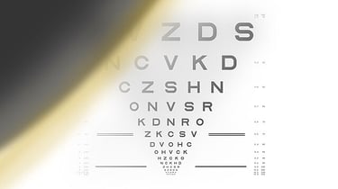
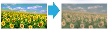
Myopia also called as nearsightedness makes distant objects appear blurry, while nearby objects remain clear. It usually begins in childhood and tends to progress over time. This common refractive error affects millions worldwide and can impact daily activities such as reading the board at school, driving, or recognizing faces from a distance. While mild myopia can often be corrected with glasses or contact lenses, progressive myopia may increase the risk of serious eye complications. Understanding how myopia develops and the factors that contribute to its progression is essential.

What causes Myopia to get worse?

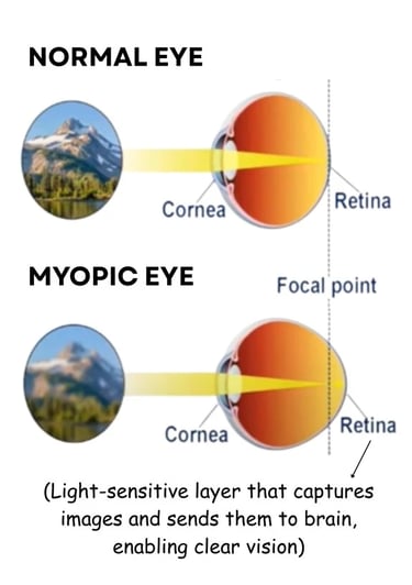
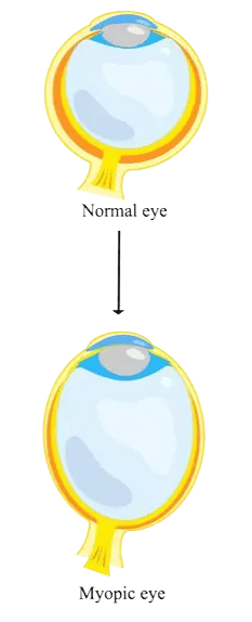
As Myopia increases, the eyeball elongates and this structural change causes light to focus in front of the retina instead of directly on it, leading to blurred distance vision. Key factors that contribute to worsening myopia include:

  • Genetic factors: Children with myopic parents are more likely to develop worsening myopia.

  • Excessive near work: Prolonged reading, screen time, or close-up tasks can speed up progression.

  • Limited outdoor time: Reduced exposure to natural light is linked to faster myopia development.

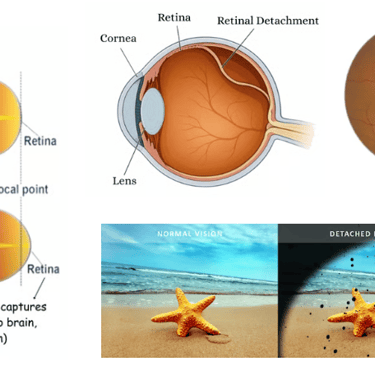
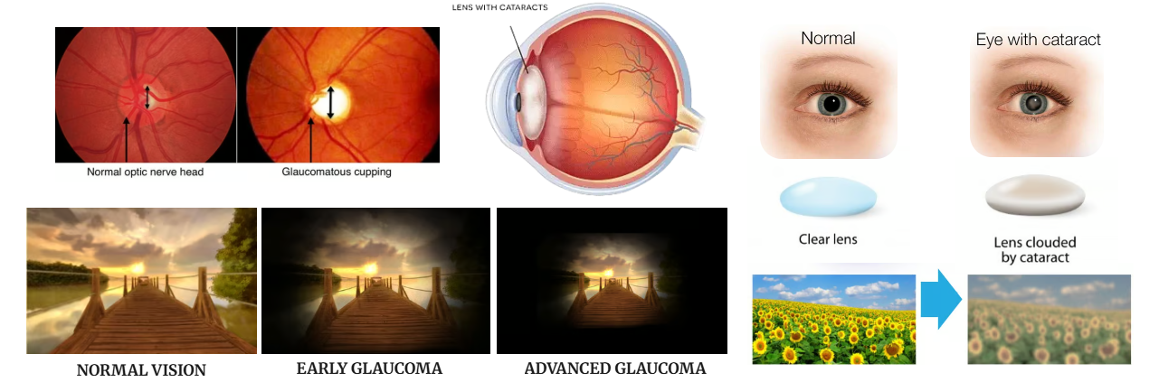
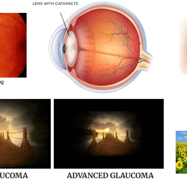
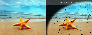
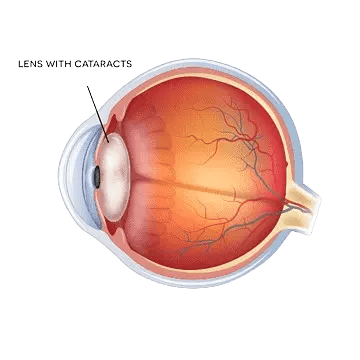
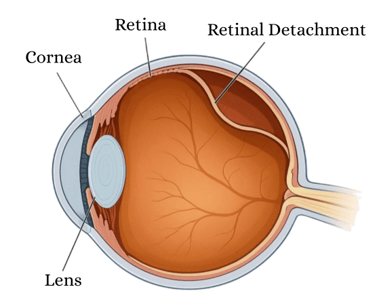
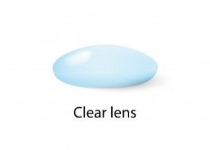
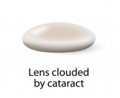
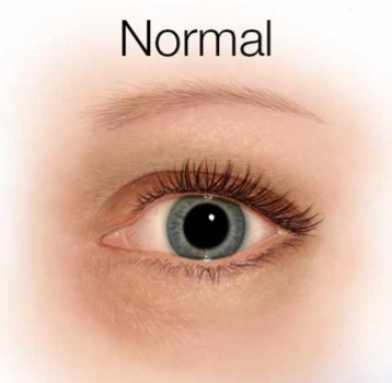
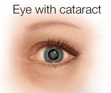
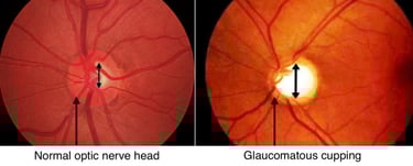
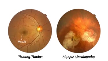
Over time, excessive elongation of the eye can lead to mechanical stretching and thinning of the retinal and choroidal tissues. This increases the risk of developing sight-threatening complications which include Retinal detachment due to peripheral retinal thinning, Myopic maculopathy caused by reduced blood supply and degeneration at the macula (center of vision), Glaucoma due to changes in optic nerve head structure and Early-onset cataracts.

The more severe myopia becomes, the greater the risk of developing these serious eye conditions, some of which can lead to irreversible vision loss . Early myopia control can help reduce the risk of long-term complications, preserve vision, and avoid the need for complex and costly treatments later in life.

Illustration of focal point of normal and myopic eye explaining about what is myopia
normal eye vs myopia eye diagram showing how myopia eye disease is caused
Normal eye vs eye with retinal detachment explaining about how vision with RD look like.
image of eye anatomy diagram showing cataractous lens
Cross-sectional diagram of an eye showing retinal detachment with cornea, lens, and retina labeled
Image of clear lens in normal person with healthy eye vs cataract lens
Image of a cloudy lens clouded with cataract.
Comparison of normal vision vs cataract vision showing field of sunflowers with cloudy cataract
Normal human eye with clear lens vs eye with cataract.
Image of human eye with cloudy cataract in lens.
How normal vision look like vs glaucoma vision illustration in scenery.
Tunnel vision image showing signs of early glaucoma in people with high eye pressure.
Tunnel vision image with advanced glaucoma in people with high IOP.
image of normal optic nerve head in healthy people vs glaucomatous cupping in eye with glaucoma
Fundus photograph showing myopic maculopathy with retinal changes near the macula

NORMAL VISION

EARLY GLAUCOMA

ADVANCED GLAUCOMA

Curtain like appearance suggesting retinal detachment.

🤔

🔍

..So Let's Explore-